Walk into any beauty clinic or skincare counter, and you’ll see a “skin analysis” device flashing data on the screen. It looks advanced, it feels scientific—but what does it really mean to the client? Is it a marketing prop or the beginning of genuine trust?
Visia-style Skin Analyzers
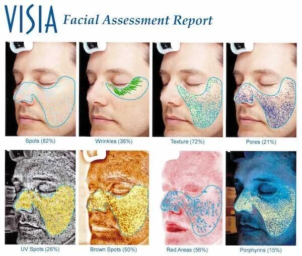
VISIA is a skin analysis system developed primarily by the American company Canfield. It is virtually the only device capable of quantitatively and comprehensively analyzing various skin features, including superficial pigmentation, deep pigmentation, vascular dilation, skin texture, pores, and sebum levels.
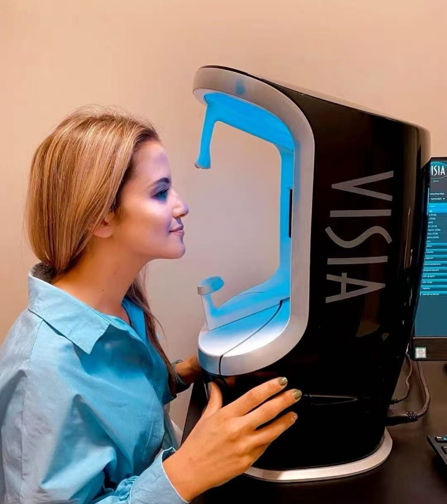
In essence, it is essentially a high-end, large-scale camera. VISIA uses standard white light, ultraviolet (UV) light, polarized light, and an adjustable xenon flash to capture images, with a resolution of up to 28 million pixels, producing a series of high-resolution photographs.
Standard white light captures the skin as seen by the naked eye, revealing the condition of the epidermis. Polarized light is used to detect surface and subsurface details, filtering out interference from external color variations, facial oil, and reflections, thereby accurately displaying the skin’s basic structure. The system can also emit a small amount of UV light to assess deeper skin conditions.
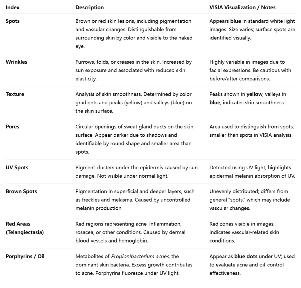
By combining these three light sources, VISIA can clearly and accurately collect information about the skin. It analyzes 8 key skin indicators: spots, wrinkles, texture, pores, UV spots, brown spots, red areas (telangiectasia), and porphyrin-related oil.
If a database is available, VISIA can also evaluate how your skin compares to the general population and provide a difference between your chronological age and your “skin age,” giving the impression of looking younger (or older). However, these results are not highly precise and should be taken with caution.
Existing literature has demonstrated that VISIA’s quantitative analysis can be effectively applied to assess conditions such as melasma, acne scars, and acne treatment outcomes. Clinically, evaluating acne or other facial skin problems still largely relies on dermatologists’ visual assessment, which is subjective and time-consuming. VISIA provides a convenient solution by capturing high-resolution images that allow direct comparison before and after treatment.
Beyond acne, VISIA can also help evaluate pigmentation, post-inflammatory hyperpigmentation, and steroid-dependent dermatitis by assessing lesion depth and observing the skin’s subsurface structure. This information can guide treatment planning and estimate treatment duration.
However, VISIA has its limitations. Its ability to evaluate skin aging is relatively limited.
Beyond the Surface: How High-Frequency Ultrasound Is Redefining Skin Diagnostics
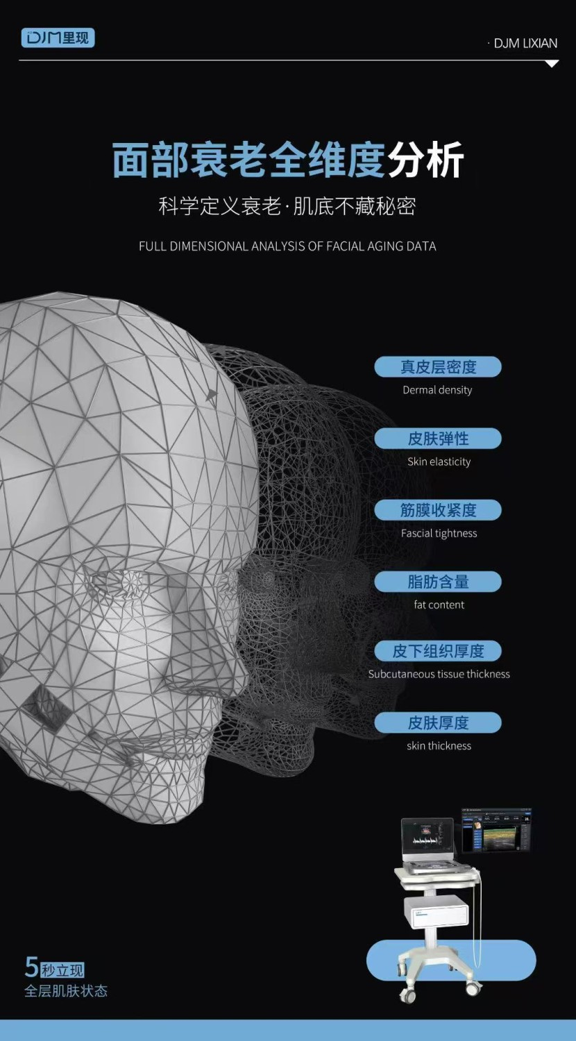
Optical imaging can only capture the skin’s surface. Yet, the most critical signs of aging and structural changes occur much deeper—within the dermis, fascia, and subcutaneous fat. This gap has created a need for deeper, more precise, and actionable skin diagnostics.
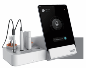
DJM Medical Instrument GmbH is pioneering the integration of high-frequency ultrasound imaging into professional skin diagnostics. This approach moves beyond optical imaging, enabling acoustic visualization and deep-layer quantitative analysis, and represents a new standard for aesthetic medicine.

Industry-Leading 19 MHz Medical-Grade Ultrasound
Our system is powered by a 19 MHz medical-grade ultrasound engine, setting a new benchmark in aesthetic diagnostics. No other device under USD 50,000 on the global market offers such high imaging frequency. This provides unmatched advantages in:
Resolution: Captures fine skin structures with exceptional clarity.
Depth penetration: Visualizes layers up to 30 mm beneath the skin surface.
Image stability: Ensures consistent, reproducible results.
- Visualization
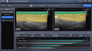
DJM allows practitioners to instantly see the skin’s real structure—epidermis, dermis, and subcutaneous layers are all clearly displayed. For instance, a forehead dermal thickness of 2.1 mm indicates healthy and resilient skin. This level of insight transforms subjective evaluation into objective understanding.
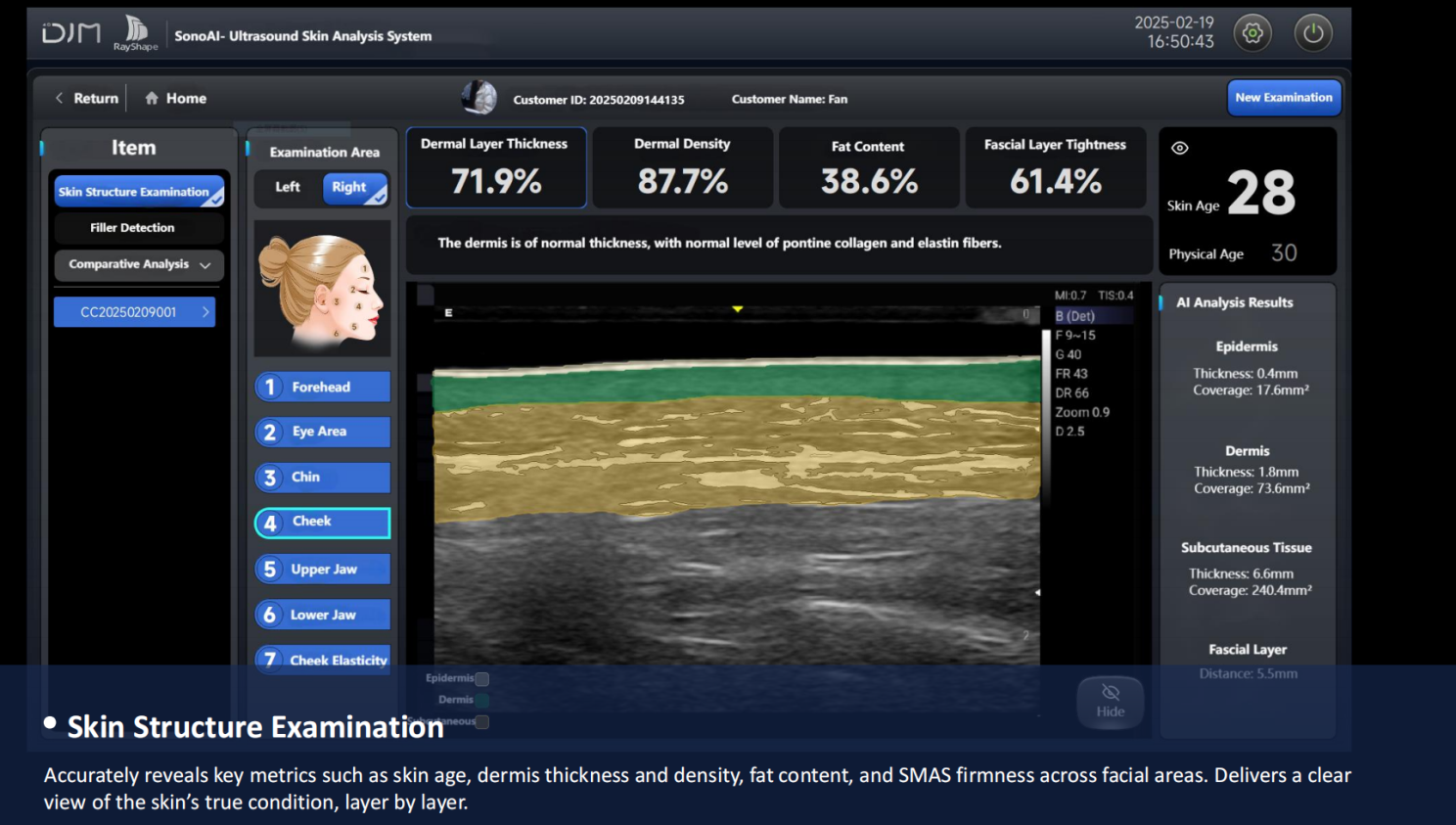
Advanced AI algorithms convert complex black-and-white ultrasound scans into intuitive, full-color maps. Even non-medical staff can interpret results accurately, improving workflow efficiency and client communication.
Backed by a database of over 18 million skin profiles, every condition is quantified and standardized, enabling consistent evaluation and long-term tracking. This data-driven approach supports personalized treatment planning and outcome prediction.
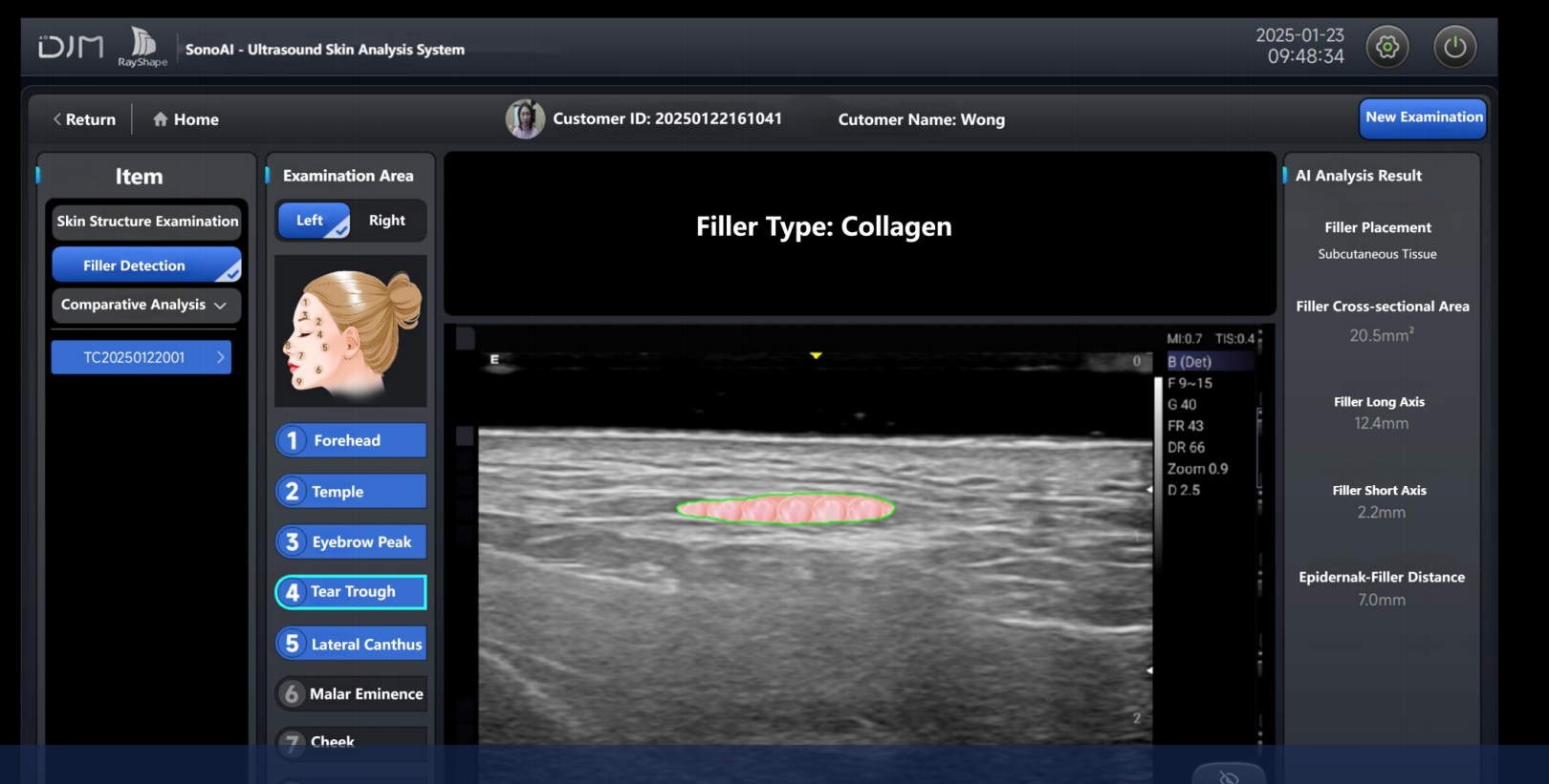
DJM can accurately detect fillers such as hyaluronic acid and collagen beneath the skin. It automatically measures filler area, long-axis, and short-axis dimensions, and identifies injection depth and layer. Previously, such procedures relied heavily on tactile experience; now, ultrasound visualization ensures precision, safety, and consistency—turning aesthetic injection from an art based on experience into a science guided by evidence.
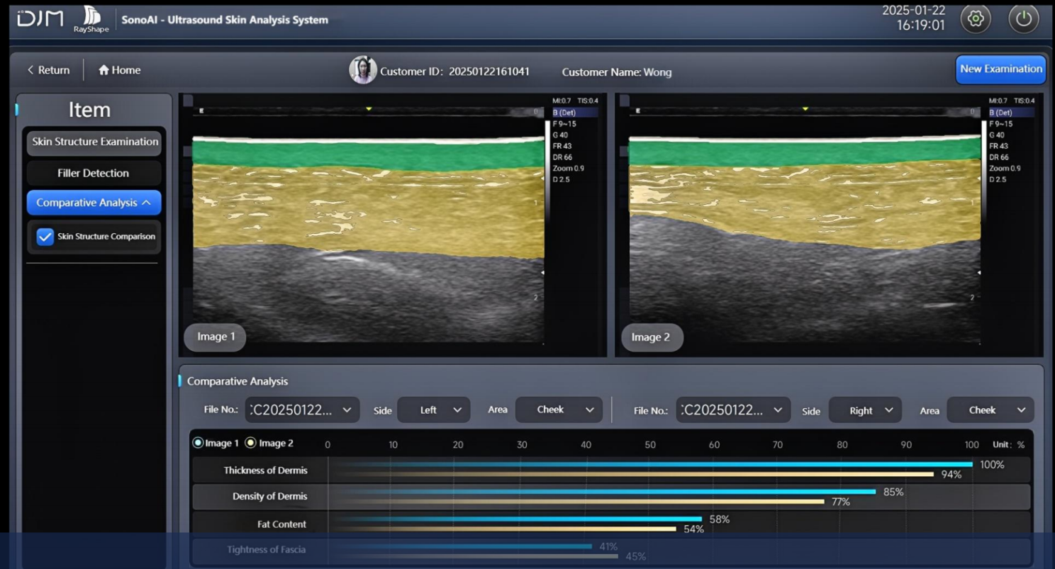
Real-Time, Transparent Procedures
The system supports real-time ultrasound visualization during injections or other treatments. Practitioners and clients can see exactly what is happening beneath the skin, ensuring transparency, reducing anxiety, and reinforcing professionalism and safety. Clients not only witness the procedure but also gain confidence in the precision and effectiveness of their treatment.


Related Products